Revista Veterinaria. 2026; 37
https://doi.org/10.30972/vet.3719486
Artículo Original


P.L. Martin1,c1
A. Koval2,c2
M.J. Butti3,c3
G. Warley1,c4
M.N. Sánchez4,c5
J.V. Petrakovsky4,c6
M.I. Gamboa3,c7
1Laboratorio de Bacteriología y Antimicrobianos (LaByAn) y Servicio Central de Laboratorio (SCL), Facultad de Ciencias Veterinarias, Universidad Nacional de La Plata (FCV- UNLP), Av. 60 y 118, CC 296, La Plata, CP 1900, Buenos Aires, Argentina.
2Biogénesis Bagó, Ruta Panamericana Km 38,5, Garin, Buenos Aires, Argentina.
3Cátedra de Parasitología Comparada-Laboratorio de Parasitosis Humanas y Zoonosis Parasitarias (LAPAHUZO), FCV-UNLP.
4Dirección Nacional de Laboratorios y Control Técnico del Servicio Nacional de Sanidad Animal (SENASA), Talcahuano 1660 Martínez, CP 1640, Buenos Aires, Argentina.
c2 ariel.koval@biogenesisbago.com
Abstract
Leptospirosis is a zoonotic disease of global importance where dogs may contribute to environmental contamination with pathogenic Leptospira. This study aimed to isolate and characterize a virulent Leptospira interrogans strain obtained from the urine of an asymptomatic dog in Argentina. Blood and urine samples were collected for hematological, biochemical, serological, and bacteriological analyses. Serology was performed using the microscopic agglutination test (MAT), and urine samples were cultured for bacterial isolation. The isolate was characterized by PCR amplification and sequencing of the rrs gene, followed by phylogenetic and serological analysis. Virulence was assessed using a susceptible hamster infection model. MAT revealed a low antibody titer (1:100) against L. interrogans serovar Canicola. Leptospira was successfully isolated from urine. Molecular and phylogenetic analyses confirmed that the isolate belonged to the pathogenic clade (P1) of L. interrogans, and serological characterization identified it as serogroup Canicola. Experimental infection in Syrian hamsters confirmed the virulence of the isolate, despite the absence of clinical signs in the dog. These findings highlight the epidemiological role of dogs as asymptomatic carriers and the importance of surveillance strategies within a One Health framework.
Key words: Leptospirosis, canine carrier, urinary shedding, zoonosis.
Resumen: La leptospirosis es una enfermedad zoonótica de importancia global en la cual los perros pueden contribuir a la contaminación ambiental con leptospiras patógenas. El objetivo de este estudio fue describir el aislamiento y la caracterización de una cepa virulenta de Leptospira interrogans obtenida a partir de la orina de un perro sin signos clínicos de leptospirosis en Argentina. Se recolectaron muestras de sangre y orina para análisis hematológicos, bioquímicos, serológicos y bacteriológicos. La serología se realizó mediante la prueba de aglutinación microscópica (MAT), y las muestras de orina fueron cultivadas para el aislamiento bacteriano. La caracterización de la cepa fue realizada mediante amplificación por PCR y secuenciación del gen rrs, seguida de análisis filogenético y caracterización serológica. La virulencia se evaluó utilizando un modelo de infección en hámsteres susceptibles. La MAT reveló un título bajo de anticuerpos (1:100) frente a L. interrogans serovariedad Canicola. Leptospiras viables fueron aisladas exitosamente a partir de la orina. Los análisis moleculares y filogenéticos confirmaron que el aislamiento pertenecía al clado patógeno (P1) de L. interrogans y la caracterización serológica lo identificó como perteneciente al serogrupo Canicola. La infección experimental en hámsteres sirios confirmó la virulencia del aislamiento, a pesar de la ausencia de signos clínicos compatibles con leptospirosis en el perro. Estos hallazgos destacan el potencial papel epidemiológico de los perros como portadores asintomáticos y la importancia de las estrategias de vigilancia dentro del enfoque Una Salud. Palabras clave: leptospirosis, portador canino, eliminación urinaria, zoonosis.
Leptospirosis is an emerging zoonosis with worldwide distribution caused by bacteria of the genus Leptospira, which currently comprises more than 64 species. Traditionally, these species are grouped into two major clades: pathogenic and saprophytic. However, within the pathogenic clade, phylogenetic analyses have identified distinct subclades, commonly referred to as P1 (pathogenic) and P2 (intermediate), reflecting different levels of virulence. This disease affects humans and numerous animal species, the latter playing an important role in its zoonotic transmission, as several mammals act as reservoirs of the microorganism (Vincent et al. 2019).
Within the 'One Health' framework (WHO), monitoring leptospirosis is particularly important as companion animals share the same habitat as humans and may therefore contribute to the environmental dissemination of Leptospira spp. In urban areas, dogs and rodents are the main species involved in environmental contamination and in the transmission of Leptospira spp., to humans. Dogs may develop either subclinical infection or severe, life-threatening disease depending on factors such as the strain virulence, infective dose, and the host´s immune status at the time of infection (Sykes et al. 2023).
In Argentina, several studies have reported exposure to Leptospira spp. in dogs, with seroprevalence varying according to geographic region and population characteristics. Rates of up to 34.9% have been described in dogs suspected of leptospirosis in the Metropolitan Area of Buenos Aires (MABA) (Martin et al. 2021). Additionally, studies conducted in MABA reported seroprevalence rates of 26.8% in dogs without clinical signs (Martin et al. 2014).
Current knowledge regarding the virulence of circulating Leptospira strains isolated from dogs in this region remains limited. The isolation of L. interrogans from the fetus of an aborted dog has been previously reported (Rossetti et al. 2005). In addition, molecular characterization of isolates obtained from dogs in La Plata and the peri-urban area of Buenos Aires revealed that all strains belonged to L. interrogans (Grune Loffler et al. 2014). All of these strains were classified as pathogenic based on molecular characterization; however, their virulence was not assessed in those studies. Therefore, the objective of the present study was to describe the isolation and characterization of a virulent L. interrogans strain obtained from the urine of a dog without clinical signs of leptospirosis in Buenos Aires Province, Argentina.
In May 2025, within the framework of a research project funded by the Programa de Incentivos Docentes, Facultad de Ciencias Veterinarias, Universidad Nacional de La Plata (FCV-UNLP; Resolution No. 11V312), a 1.5-year-old intact male mixed-breed dog from the El Molino neighborhood, Ensenada, Buenos Aires Province, was examined during a routine visit for annual rabies vaccination and primary health care. The study was performed following international recommendations specified in the guidelines for the care and use of laboratory animals and the National Academy of Sciences recommendations concerning the use of dogs as laboratory animals (Council for International Organizations of Medical Sciences International Guiding Principles for Biomedical Research Involving Animals, WHO), and the approval of the Comité Institucional para el Cuidado y Uso de Animales de Laboratorio (CICUAL) of the Facultad de Ciencias Veterinarias, Universidad Nacional de La Plata (approval no. 231017-8). Biological samples were collected by the attending veterinarian with the prior informed consent of the animal owner.
A complete physical examination was performed during the visit. Blood samples were collected with anticoagulants for complete blood count (CBC) analysis and without anticoagulants for serum biochemistry and leptospirosis serology. Abdominal ultrasonography was conducted, and urine samples were collected by catheterization for culture and bacterial isolation. The CBC was performed using both automated and manual methods. Automated analyses were conducted using a Sysmex® hematology analyzer, while manual procedures included hematocrit determination and differential leukocyte counts. Serum biochemical analyses were performed using an INCCA® analyzer.
The microscopic agglutination test (MAT) was performed according to the standard protocol. Serum samples were tested using two-fold dilutions from 1:100 to 1:6400 against a panel of reference Leptospira serovars including Castellonis, Canicola, Copenhageni, Pomona, Hardjo, Wolffi, Grippotyphosa, Pyrogenes, and Tarassovi. The endpoint titer was defined as the highest serum dilution showing ≥50% agglutination of leptospires. Titers ≥1:100 were considered positive.
For culture and isolation, urine samples were processed within two hours of collection. A 100 µL aliquot was inoculated into 5 mL of semisolid Ellinghausen-McCullough-Johnson-Harris (EMJH) medium supplemented with 100 µg mL-1 of 5-fluorouracil (5-FU; Sigma). Three serial 1:50 dilutions were subsequently prepared from the initial inoculated tube into fresh EMJH medium containing 5-FU. All cultures were incubated at 29 °C and examined weekly for the presence of spirochetes.
Serological and molecular methods were used for strain characterization. Genus and pathogenic clade identification were performed by PCR amplification of fragments of the rrs and lipL32 genes, following previously described protocols (Merien et al. 2005, Stoddard et al. 2009). Species identification was performed by PCR amplification and sequencing of a partial fragment of the rrs gene, following the approach described by Cilia et al. (2021). The amplified product was purified using a Wizard SV Gel and PCR Clean-Up System kit (Promega) and subjected to Sanger sequencing at the Centro de Excelencia en Medicina Traslacional (CEMET), Hospital El Cruce, Buenos Aires, Argentina. The resulting sequence was aligned with reference sequences using the ClustalW algorithm (Larkin et al. 2007). The consensus sequence was deposited in GenBank (accession number PX497584). Phylogenetic reconstruction was performed using the Neighbor-Joining method implemented in MEGA version 11 (Tamura et al. 2021), with evolutionary distances calculated using the Maximum Composite Likelihood method. Ambiguous positions were removed using the pairwise deletion option, resulting in a final dataset of 460 positions.
Serogroup identification was performed at the reference laboratory of the Servicio Nacional de Sanidad y Calidad Agroalimentaria (SENASA), Martínez, Buenos Aires, Argentina, using the MAT with a panel of specific rabbit antisera covering 24 Leptospira serogroups (Icterohaemorrhagiae, Canicola, Pomona, Sejroe, Grippotyphosa, Australis, Autumnalis, Ballum, Bataviae, Hebdomadis, Javanica, Panama, Pyrogenes, Tarassovi, Cynopteri, Mini, Shermani, Louisiana, Celledoni, Hurstbridge, Djasiman, Sarmin, Semaranga, Andamana).
Virulence assessment of the isolate was performed at the same reference laboratory through experimental infection of susceptible animals, in accordance with national regulations on animal welfare and biosafety and under the institutional authorization of SENASA. An experimental infection model using golden Syrian hamsters (Mesocricetus auratus) was employed. Five young hamsters (approximately 50-90 g) were inoculated intraperitoneally with 0.5 mL of a culture suspension of the isolate grown in EMJH medium, containing approximately 10⁸ leptospires mL-1. Animals were monitored daily for clinical signs compatible with leptospirosis and mortality for 14 days post-inoculation. Virulence was determined by the occurrence of clinical disease and/or death during the observation period, consistent with the standard hamster model for pathogenic Leptospira. Animals that died during the experiment were subjected to bacteriological confirmation by direct immunofluorescence (DIF) performed on kidney tissue.
The patient’s medical history revealed a prior diagnosis of canine distemper, parasitic infestations, and the absence of vaccination against Leptospira spp. Physical examination showed the dog to be alert, with a body condition score (BCS) of 3/5, a rough hair coat, and neurological sequelae consistent with distemper. CBC results were as follows (reference ranges in parentheses): hematocrit 30% (37-55%) red blood cells 4,800,000 µL-1 (5,500,000-8,500,000), hemoglobin 10.1 g dL-1 (12-18), mean corpuscular hemoglobin (MCH) 21.04 pg (19.5-24.5), mean corpuscular volume (MCV) 62.50 fL (60-77), mean corpuscular hemoglobin concentration (MCHC) 33.67% (32-36), platelets 369,000 µL-1 (160,000-430,000), and leukocytes 22,300 µL-1 (6,000-17,000). Differential leukocyte counts included band neutrophils 0 (<510), segmented neutrophils 16,279 µL-1 (3,600-13,100), eosinophils 2,676 µL-1 (120-1,700), basophils 0 µL-1 (0-150) lymphocytes 3,345/µL (700-5,100), and monocytes 0/µL (180-1,700). The serum biochemical profile revealed glucose 68 mg dL-1 (70-115), urea 60 mg dL-1 (20-40), creatinine 0.9 mg dL-1 (<1.5), alanine transaminase (ALT) 26 U L-1 (<66), total protein 6.56 g dL-1 (5.3-7.8), albumin 2.7 g dL-1 (2.3-4.3), globulins 3.86 g dL-1 (3.1-4.6), and an albumin/globulin ratio of 0.70. Abdominal ultrasonography revealed multiple hyperechoic structures with echogenic borders and an anechoic center, approximately 7 mm in diameter, in longitudinal and transverse sections, consistent with Dioctophyme renale.
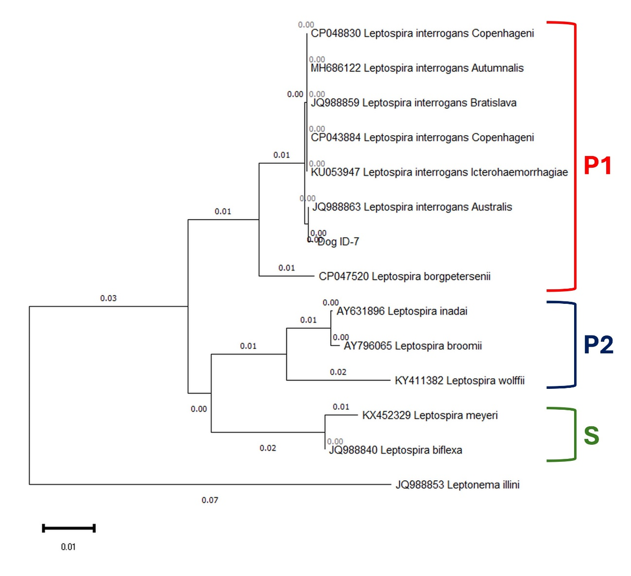
MAT revealed a titer of 1:100 against L. interrogans serovar Canicola, with no reactivity to the other serovars tested. After nine weeks of incubation, spirochetes were observed in the second tube of the dilution series. PCR amplification of rrs and lipL32 gene fragments confirmed the isolate as belonging to the genus Leptospira and to the pathogenic clade I. Phylogenetic analysis based on the 541 bp rrs gene sequence of the isolate (Dog ID-7) revealed clustering within the pathogenic clade of Leptospira, specifically in subclade P1, and clearly separated from subclade P2 (intermediate) within the pathogenic group, as well as from the saprophytic clade (S) (Figure 1).
The isolate showed a close phylogenetic relationship with reference strains of L. interrogans. Leptonema illini was included as an outgroup to root the tree. MAT with specific antisera identified the strain as belonging to the Canicola serogroup. Virulence of the isolate was confirmed using the Syrian hamster infection model described above.
Different Leptospira serovars are maintained in the environment by multiple animal species acting as hosts, thereby promoting bacterial persistence and transmission to other animals and humans. In this context, identifying both the host species and the infecting strains is essential for a comprehensive assessment of health risks (Harran et al. 2024).
The present study describes the isolation, molecular and serological characterization of a pathogenic and virulent Leptospira strain from the urine of a dog without clinical signs of leptospirosis in Buenos Aires, Argentina. Previous studies in this region have documented exposure to leptospires in dogs through serological testing and bacterial isolation. However, information regarding the immune status of these animals at the time of isolation, as well as the virulence of circulating strains, remains limited (Grune Loffler et al. 2014, Martin et al. 2014).
Regarding clinical presentation, subclinical infection has been proposed as the most common form of leptospirosis in dogs (Sykes et al. 2023). This was also observed in the present case, as no clinical signs compatible with leptospirosis were detected. These findings are consistent with previous reports describing the identification of L. interrogans serovar Canicola in dogs without compatible clinical signs (Piredda et al. 2022, Carmona Gasca et al. 2024). In contrast to some reports, there was no documented history of vaccination against leptospirosis in the dog described in the present study.
Laboratory abnormalities most frequently reported in canine leptospirosis include leukocytosis, anemia, thrombocytopenia, azotemia, and increased activities of transaminases and alkaline phosphatase (Sykes et al. 2023). In the present case, the dog exhibited mild anemia, mature neutrophilic leukocytosis, and a slight increase in serum urea concentration, despite the absence of clinical manifestations compatible with leptospirosis. This pattern differs from that described in previous reports, in which increased transaminase and alkaline phosphatase activities were the only laboratory abnormalities observed (Piredda et al. 2022). However, given the presence of concurrent conditions, including D. renale infection, the clinicopathological alterations observed in this animal cannot be conclusively attributed to leptospiral infection (Shaban et al. 2025). In addition, the dog presented neurological signs consistent with possible sequelae of canine distemper, such as myoclonus, although infection with canine distemper virus had not been confirmed. This infection has been associated with transient or prolonged immunosuppression, which may affect both humoral and cellular immune responses. If present, this condition could potentially influence the host’s ability to mount an adequate antibody response against Leptospira, which might partly explain the low MAT titer observed. Therefore, these concurrent conditions should be considered potential confounding factors when interpreting the clinical and laboratory findings in this case.
On the other hand, the detection of low antibody titers by MAT in conjunction with successful bacterial isolation is consistent with previous studies reporting molecular or culture-based detection of Leptospira in the absence of significant serological reactivity (Martin et al. 2019, Piredda et al. 2022). This apparent discrepancy underscores the complexity of leptospirosis diagnosis and highlights the importance of combining serological, molecular, and bacteriological methods for the identification of infected animals and asymptomatic carriers. Furthermore, the present findings indicate that active bacterial shedding in urine (leptospiruria) should not be excluded solely on the basis of negative or low-titer MAT results.
The molecular and serological characterization of the isolate obtained in this study is consistent with the epidemiological context of the region. Previous reports have described a predominant circulation of the Canicola serogroup of L. interrogans in canine urine samples, while the Pomona and Icterohaemorrhagiae serogroups were detected at lower frequencies (Grune Loffler et al. 2014, Carmona Gasca et al. 2024). Similarly, molecular characterisation of isolates obtained from human cases of leptospirosis in Argentina has identified L. interrogans serogroup Canicola as the predominant genotype (Chiani et al. 2016). These findings, together with previous reports from the region, suggest that strains belonging to the Canicola serogroup may circulate among canine populations and that dogs may contribute to the maintenance and dissemination of pathogenic Leptospira strains in urban and peri-urban environments.
A limitation of the present report is that it describes a single clinical case; therefore, broader epidemiological inference is limited. In addition, species identification and phylogenetic inference were based on a partial fragment of the rrs gene. Although this marker is widely used for the genus- and species-level identification of Leptospira, it provides limited resolution for strain typing compared with multilocus sequence typing (MLST) or whole-genome sequencing approaches. Future studies incorporating MLST or genomic analyses may allow a more detailed characterization of circulating strains.
This report describes the isolation and characterization of a virulent L. interrogans strain belonging to the Canicola serogroup from the urine of a dog without clinical signs in Buenos Aires, Argentina. Although based on a single case, this finding demonstrates that dogs without apparent clinical disease may shed pathogenic leptospires in urine. These observations highlight the potential epidemiological relevance of asymptomatic canine carriers and reinforce the importance of integrated surveillance strategies combining bacteriological, molecular, and serological approaches for the detection of circulating Leptospira strains in companion animals within a One Health framework.
Acknowledgment.The authors gratefully acknowledge the valuable technical assistance provided by Keila Zegarra Borlando (SCL) and Fabio Nievas (LaByAn).
Author Contributions. MPL and MIG contributed to the development and initial drafting of the manuscript. MPL, KA, BMJ and WG were responsible for sample processing, diagnostic testing, molecular and phylogenetic analysis of the samples. SM and PJ were responsible for serological characterization and virulence determination. All authors contributed to the editing and review of the manuscript. Furthermore, all authors approved the final version for submission.
Conflict of interest. The authors declare that they have no known financial conflicts of interest or personal relationships that could have influenced the work reported in this article.